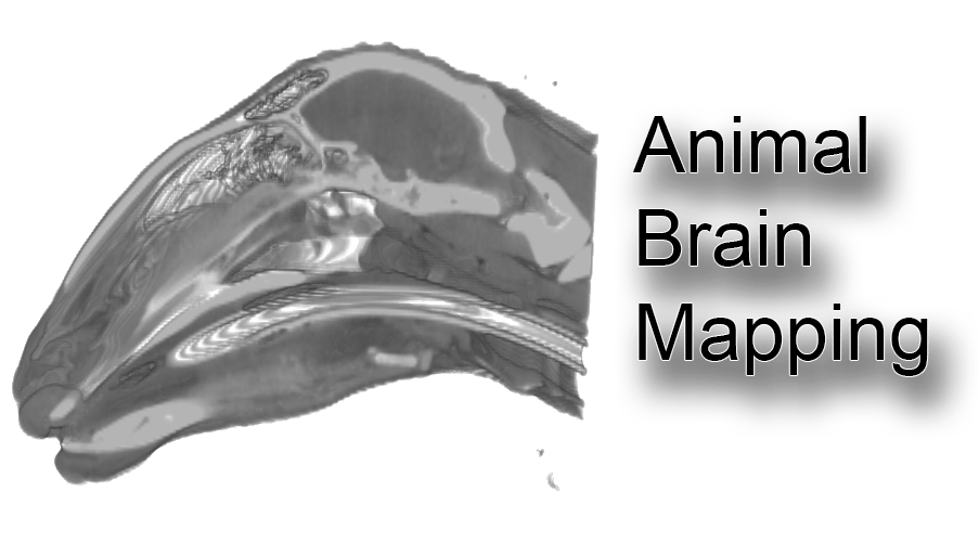
OBJECTIVES: We present an unbiased, population-averaged standard magnetic resonance imaging (MRI) template of ovine brain volume. This template acts as a common stereotaxic reference frame for the accurate localization of anatomical and functional information, enabling organized comparisons across individual sheep and various studies. Utilizing T1-weighted MRI volumes from a cohort of 14 normal adult sheep, we generated the template along with prior probabilities for cerebral gray matter (GM), white matter (WM), and cerebrospinal fluid (CSF). Consequently, this atlas is derived not from the anatomy of a single subject but through the nonlinear normalization of multiple sheep brains, which are mapped to an average template image that accurately represents the locations of anatomical structures.
METHODS: The ovine average atlas consists of T1-weighted MRIs from 14 normal young adult sheep brains (Ovis orientalis aries). This atlas is constructed from an average that accounts for each individual's position, orientation, and scale, ensuring it accurately reflects both the intensities and spatial arrangement of anatomical structures. Image preprocessing involved non-uniform intensity correction and intensity normalization within a range of 0 to 100. One sheep was designated as the initial target, and the registration of the remaining 13 sheep brains commenced using manually identified homologous landmarks. These landmarks included the centers of the left and right eyeballs, the most rostral and caudal poles of both hemispheres, the intersection of the cruciate sulcus and the longitudinal fissure, as well as the anterior (AC) and posterior commissures (PC).
A three-step average template generation process (Fonov et al., 2009) was employed to estimate the nonlinear deformation field that best aligned local neighborhoods between each sheep brain volume and the average target. In brief, transformation matrices calculated in one step were applied to the corresponding scans for image resampling, which were subsequently utilized in the following step. The processing included: first, a linear left-right symmetric rigid body registration using eight iterations to create a rigidly transformed average. Next, a left-right symmetric fully-affine (12 parameters) iterative registration procedure was conducted on each input scan, followed by the calculation of the average over eight iterations. Finally, a left-right symmetric nonlinear registration was performed involving four iterations with 4 mm steps, followed by four iterations with 2 mm steps, and four iterations with 1 mm steps, culminating in the creation of the final nonlinear transformed average.
PUBLICATIONS: The following publications need to be cited when utilizing this atlas:
Nitzsche B, Frey S, Collins LD, Seeger J, Lobsien D, Dreyer A, Kirsten H, Stoffel MH, Fonov VS, and Boltze J (2015). A stereotaxic, population-averaged T1w ovine brain atlas including cerebral morphology and tissue volumes. *Frontiers in Neuroanatomy*, 9:69. doi: 10.3389/fnana.2015.00069
Fonov V, Evans AC, Botteron K, Almli CR, McKinstry RC, and Collins DL (2011). Unbiased average age-appropriate atlases for pediatric studies. *Neuroimage*, 54(1), 313–327.
LICENSE: The dataset is licensed under CC BY International 4.0. You are free to share and adapt the dataset, but you must provide appropriate credit, link to the license, and indicate if changes were made (see more [here]).
[Link to McGill-Neuroinformatic]
DOWNLOADS: see below
OBJECTIVE: The presented atlas labels are derived from refined gray/white matter masks of the T1-weighted template, ensuring strict alignment with both the template and the tissue probability maps (TPM).
METHODS: The gray/white matter masks were delineated manually and semi-automatically using FIJI in orthogonal view. Results were iteratively validated using MRIcro. The data were tested during the processing of Positron Emission Tomography (PET) data in a ovine Huntington's disease model .
PUBLICATION: Please cite the following publication when using the atlas labels in your own research:
Williams GK, Akkermans J, Lawson M, Syta P, Staelens S, Adhikari MH, Morton AJ, Nitzsche B, Boltze J, Christou C, Bertoglio D, Ahamed M. Imaging Glucose Metabolism and Dopaminergic Dysfunction in Sheep (Ovis aries) Brain Using Positron Emission Tomography Imaging Reveals Abnormalities in OVT73 Huntington's Disease Sheep. ACS Chem Neurosci. 2024 Nov 6;15(21):4082-4091. doi: 10.1021/acschemneuro.4c00561. Epub 2024 Oct 17. PMID: 39420554.
LICENCE: The dataset is licensed under CC BY International 4.0 . You are free to share and adapt the data set, but required to give appropriate credit, provide a link to the license and indicate if changes were made (see more [here]).